ファイル:Surface projections of the organs of the trunk.png

ページのコンテンツが他言語でサポートされていません。
出典: フリー教科書『ウィキブックス(Wikibooks)』

元のファイル(1,583 × 2,533 ピクセル、ファイルサイズ: 3.33メガバイト、MIME タイプ: image/png)

このファイルはウィキメディア・コモンズから来ており、他のプロジェクトで使用されている可能性があります。 ウィキメディア・コモンズでのファイル解説ページにある説明を以下に示します。

概要

解説
English: Surface projections of the major organs of the trunk, using the vertebral column and rib cage as main reference points of superficial anatomy. The transpyloric plane and McBurney's point are among the marked locations.

To discuss image, please see Talk:Human body diagrams

日付
原典

All included images are in public domain. See Template:Human body diagrams for individual details.

References

Spinal vertebrae levels:

  • Located at middle of frontal face of spinal vertebrae according to reference the image :File:Gray 111 - Vertebral column.png when pasted into image to superiorly fit the surface bulges of the clavicular bones and inferiorly fit the surface bulges of the anterior iliac spine.

Gross overview of organ locations:

  • Plate 260B - Regions and Planes of Adbomen. From: The Interactive Atlas of Human Anatomy Version 3.0 (CD-ROM). Authors: Frank H. Netter and Carlos A. G. Machado. Published by Saunders, October 1, 2002. ISBN-10: 1929007140. ISBN-13: 978-1929007141.
  • Fig 139 from The SPENCER SYSTEM. SEVENTEENTH EDITION
  • Plate 1034 from Gray's Anatomy

Locations of specific organs:
The following vertebral levels are generally given by the middle of the vertebral body.

  • Bålens ytanatomy (Superficial anatomy of the trunk). Anca Dragomir, Mats Hjortberg and Godfried M. Romans. Section for human anatomy at the Department of medical cell biology, Uppsala university, Sweden.
Key points from that compendium:
  • The oblique fissure of the right lung goes from the intercostal space 4 to 6
  • The horizontal fissure goes along the 4th rib (the original image from Gray has this line almost at the 5th rib, but this is not labeled in image, as other lung images seem to support the upper level)
  • The cardia of the stomach is at the level of the 10th rib
  • The fundus of the stomach is in the 5th intercostal space, slightly below the apex of the heart
  • Pylorus is usually at the L1 (the transpyloric plane)
  • The transpyloric plane is at the level of:
    • lumbar vertebra 1
    • the pancreatic body
    • the origins of the superior mesenteric artery from the aorta and portal vein
    • the left and right colic flexure
    • hilum of the kidney on the left
    • upper pole of the kidney on the right
    • duodenojejunal flexure
  • McBurney's point is located one third of a line from the anterior superior iliac spine and the navel
  • The upper rim of the liver is in the 5th intercostal space
  • The left lobe of the liver is to the left of the midline
  • The lower limit of the liver at the midline is approximately 3 cm below processus xiphoideus
  • The spleen is projected against the 9th and 10th rib on the backside
  • The left kidney is approximately at the vertebral level T12 to L3, and the right one slightly lower.
  • Atlas of Human Anatomy in Cross Section: Appendix: Topography of the Thorax and Abdomen. Ronald A. Bergman, Ph.D., Adel K. Afifi, M.D., Jean J. Jew, M.D., and Paul C. Reimann, B.S. [1]:
  • The average position of the highest point of the spleen is at the level of the lower third of the tenth thoracic vertebra.
  • The average position of the lowest point of the spleen is at the level of the middle third of the first lumbar vertebra.

Position of the navel

Urinary tract:

作者 Mikael Häggström
このファイルのベクター画像 (SVG) が利用できます。 使う目的に対し、元画像よりもSVGがより優れている場合、SVG画像を使用して下さい。

File:Surface projections of the organs of the trunk.png → File:Surface projections of the organs of the trunk.svg

ベクターグラフィックスの詳細については、コモンズのSVG変換を参照ください。
SVGのMediaWikiのサポートに関する情報もあります。

他言語
Alemannisch  Bahasa Indonesia  Bahasa Melayu  British English  català  čeština  dansk  Deutsch  eesti  English  español  Esperanto  euskara  français  Frysk  galego  hrvatski  Ido  italiano  lietuvių  magyar  Nederlands  norsk bokmål  norsk nynorsk  occitan  Plattdüütsch  polski  português  português do Brasil  română  Scots  sicilianu  slovenčina  slovenščina  suomi  svenska  Tiếng Việt  Türkçe  vèneto  Ελληνικά  беларуская (тарашкевіца)  български  македонски  нохчийн  русский  српски / srpski  татарча/tatarça  українська  ქართული  հայերեն  বাংলা  தமிழ்  മലയാളം  ไทย  한국어  日本語  简体中文  繁體中文  עברית  العربية  فارسی  +/−
新しいSVG画像

ライセンス

Public domain この著作物の著作権者である私は、この著作物における権利を放棄しパブリックドメインとします。これは全世界で適用されます。
一部の国では、これが法的に可能ではない場合があります。その場合は、次のように宣言します。
私は、あらゆる人に対して、法により必要とされている条件を除き、如何なる条件も課すことなく、あらゆる目的のためにこの著作物を使用する権利を与えます。

Human body diagrams

Main article at: Human body diagrams

Template location:Template:Human body diagrams

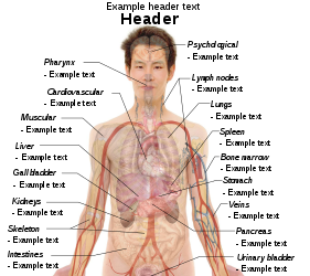
How to derive an image

Derive directly from raster image with organs

The raster (.png format) images below have most commonly used organs already included, and text and lines can be added in almost any graphics editor. This is the easiest method, but does not leave any room for customizing what organs are shown.

Adding text and lines:

Derive "from scratch"

By this method, body diagrams can be derived by pasting organs into one of the "plain" body images shown below. This method requires a graphics editor that can handle transparent images, in order to avoid white squares around the organs when pasting onto the body image. Pictures of organs are found on the project's main page. These were originally adapted to fit the male shadow/silhouette.

Organs:

More organs are found at: Human body diagrams/Organs

Derive by vector template

The Vector templates below can be used to derive images with, for example, Inkscape. This is the method with the greatest potential.

See Human body diagrams/Inkscape tutorial for a basic description in how to do this.

Examples of derived works

More examples

Licensing

Creative Commons CC-Zero このファイルはクリエイティブ・コモンズ CC0 1.0 全世界 パブリック・ドメイン提供のもとで利用可能にされています。
ある作品に本コモンズ証を関連づけた者は、その作品について世界全地域において著作権法上認められる、その者が持つすべての権利(その作品に関する権利や隣接する権利を含む。)を、法令上認められる最大限の範囲で放棄して、パブリック・ドメインに提供しています。

この作品は、たとえ営利目的であっても、許可を得ずに複製、改変・翻案、配布、上演・演奏することが出来ます。

キャプション

このファイルの内容を1行で記述してください

このファイルに描写されている項目

題材

5 9 2010

ファイルの履歴

過去の版のファイルを表示するには、その版の日時をクリックしてください。

(最新 | 最古) (以後の10件 | ) (10 | 20 | 50 | 100 | 250 | 500 件) を表示
日付と時刻サムネイル寸法利用者コメント
現在の版2019年12月27日 (金) 09:192019年12月27日 (金) 09:19時点における版のサムネイル1,583 × 2,533 (3.33メガバイト)Mikael Häggström+Costal margin
2010年11月11日 (木) 10:382010年11月11日 (木) 10:38時点における版のサムネイル1,050 × 1,680 (2.07メガバイト)Mikael HäggströmAdapted to recently added overview images. Distinguished different ways to designate vertebrae levels.
2010年11月7日 (日) 10:042010年11月7日 (日) 10:04時点における版のサムネイル936 × 1,325 (1.77メガバイト)Mikael Häggströmupdate from svg
2010年11月7日 (日) 09:462010年11月7日 (日) 09:46時点における版のサムネイル936 × 1,325 (1.77メガバイト)Mikael Häggströmupdate from svg
2010年10月24日 (日) 04:512010年10月24日 (日) 04:51時点における版のサムネイル936 × 1,325 (1.61メガバイト)Mikael HäggströmSmoother edges
2010年10月10日 (日) 05:182010年10月10日 (日) 05:18時点における版のサムネイル936 × 1,325 (1.61メガバイト)Mikael HäggströmMinor kidney adjustment. More realistic hip bone
2010年10月6日 (水) 04:472010年10月6日 (水) 04:47時点における版のサムネイル936 × 1,325 (1.73メガバイト)Mikael HäggströmDistinguished stomach and spleen. Removed painted arteries out of scope.
2010年10月4日 (月) 18:402010年10月4日 (月) 18:40時点における版のサムネイル936 × 1,325 (1.74メガバイト)Mikael HäggströmLowered spleen
2010年10月3日 (日) 15:212010年10月3日 (日) 15:21時点における版のサムネイル936 × 1,325 (1.74メガバイト)Mikael HäggströmDecreased some opacity. Aligned tail of pancreas with spleen. Adjusted fissure marking width.
2010年10月2日 (土) 18:202010年10月2日 (土) 18:20時点における版のサムネイル936 × 1,325 (1.72メガバイト)Mikael Häggström+liver label
(最新 | 最古) (以後の10件 | ) (10 | 20 | 50 | 100 | 250 | 500 件) を表示

グローバルなファイル使用状況

以下に挙げる他のウィキがこの画像を使っています:

このファイルのグローバル使用状況を表示する。

メタデータ